If You Have Already Have Had a Ovarian Cyst Can You Get One Again
| Ovarian cyst | |
|---|---|
 | |
| A simple ovarian cyst of most likely follicular origin | |
| Specialty | Gynecology |
| Symptoms | None, bloating, lower abdominal pain, lower dorsum hurting[1] |
| Complications | Rupture, twisting of the ovary[1] |
| Types | Follicular cyst, corpus luteum cyst, cysts due to endometriosis, dermoid cyst, cystadenoma, ovarian cancer[1] |
| Diagnostic method | Ultrasound[1] |
| Prevention | Hormonal nascency control[1] |
| Treatment | Conservative management, hurting medication, surgery[1] |
| Prognosis | Ordinarily good[1] |
| Frequency | 8% symptomatic before menopause[ane] |
An ovarian cyst is a fluid-filled sac inside the ovary.[ane] Often they cause no symptoms.[ane] Occasionally they may produce bloating, lower abdominal pain, or lower back pain.[ane] The majority of cysts are harmless.[i] If the cyst either breaks open up or causes twisting of the ovary, it may cause severe pain.[1] This may event in vomiting or feeling faint,[1] and even cause head aches.
Nearly ovarian cysts are related to ovulation, being either follicular cysts or corpus luteum cysts.[1] Other types include cysts due to endometriosis, dermoid cysts, and cystadenomas.[i] Many small cysts occur in both ovaries in polycystic ovary syndrome (PCOS).[i] Pelvic inflammatory affliction may also result in cysts.[one] Rarely, cysts may be a grade of ovarian cancer.[1] Diagnosis is undertaken by pelvic examination with an ultrasound or other testing used to gather farther details.[one]
Oft, cysts are merely observed over fourth dimension.[i] If they cause pain, medications such as paracetamol (acetaminophen) or ibuprofen may be used.[1] Hormonal birth control may be used to prevent further cysts in those who are frequently affected.[1] Notwithstanding, bear witness does not support nativity control as a handling of electric current cysts.[2] If they practise not go away after several months, get larger, look unusual, or crusade pain, they may be removed by surgery.[1]
Most women of reproductive age develop pocket-sized cysts each month.[1] Large cysts that cause problems occur in nigh 8% of women before menopause.[1] Ovarian cysts are present in almost 16% of women after menopause and if present are more than likely to be cancer.[1] [3]
Signs and symptoms [edit]

Image of multiple ovarian cysts.
Some or all of the following symptoms may be present, though it is possible not to experience any symptoms:[4]
- Abdominal pain. Deadening aching pain within the abdomen or pelvis, peculiarly during intercourse.
- Uterine bleeding. Hurting during or shortly after starting time or end of menstrual period; irregular periods, or aberrant uterine bleeding or spotting.
- Fullness, heaviness, pressure, swelling, or bloating in the abdomen.
- When a cyst ruptures from the ovary, in that location may be sudden and abrupt pain in the lower belly on ane side.
- Change in frequency or ease of urination (such as inability to fully empty the bladder), or difficulty with bowel movements due to pressure on adjacent pelvic anatomy.
- Constitutional symptoms such as fatigue, headaches.
- Nausea or vomiting
- Weight gain
Other symptoms may depend on the crusade of the cysts:[4]
- Symptoms that may occur if the cause of the cysts is polycystic ovarian syndrome (PCOS) may include increased facial hair or body hair, acne, obesity and infertility.
- If the cause is endometriosis, and so periods may be heavy, and intercourse painful.
The outcome of cysts not related to PCOS on fertility is unclear.[5]
Cyst rupture [edit]
A ruptured ovarian cyst is usually self-limiting, and simply requires keeping an eye on the situation and pain medications. The main symptom is intestinal pain, which may last a few days to several weeks, just they can also be asymptomatic.[6] Rupture of large ovarian cysts tin can cause bleeding within the abdominal cavity and in some cases shock.[7]
Ovarian torsion [edit]
Ovarian cysts increase the risk for ovarian torsion; cysts which are larger than 4 cm are associated with approximately 17% risk. The torsion tin cause obstacle of blood flow and pb to infarction.[viii]
Types [edit]
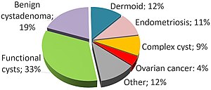
Relative incidences of dissimilar types of ovarian cysts.[nine]
Functional [edit]
Functional cysts form equally a normal part of the menstrual wheel. At that place are several types of functional cysts:
- Follicular cyst, the about common blazon of ovarian cyst. In menstruating women, a follicle containing the ovum, an unfertilized egg, volition rupture during ovulation. If this does not occur, a follicular cyst of more than 2.5 cm diameter may result.[4]
- Corpus luteum cysts announced after ovulation. The corpus luteum is the remnant of the follicle afterwards the ovum has moved to the fallopian tubes. This usually degrades within 5 to 9 days. A corpus luteum that is more than than 3 cm is defined equally cystic.[4]
- Theca lutein cysts occur within the thecal layer of cells surrounding developing oocytes. Under the influence of excessive hCG, thecal cells may proliferate and become cystic. This is usually on both ovaries.[4]
Non-functional [edit]
Non-functional cysts may include the post-obit:[ commendation needed ]
- An ovary with many cysts, which may be found in normal women, or within the setting of polycystic ovary syndrome
- Cysts caused by endometriosis, known as chocolate cysts
- Hemorrhagic ovarian cyst
- Dermoid cyst
- Ovarian serous cystadenoma
- Ovarian mucinous cystadenoma
- Paraovarian cyst
- Cystic adenofibroma
- Borderline tumoral cysts
Diagnosis [edit]

A 2 cm left ovarian cyst as seen on ultrasound

4 kinds of ovarian cysts on MRI
Ovarian cysts are normally diagnosed by ultrasound, CT scan, or MRI, and correlated with clinical presentation and endocrinologic tests as appropriate.[10]
Ultrasound [edit]
Follow-up imaging in women of reproductive age for incidentally discovered simple cysts on ultrasound is not needed until 5 cm, as these are usually normal ovarian follicles. Simple cysts five to 7 cm in premenopausal females should be followed yearly. Simple cysts larger than seven cm require further imaging with MRI or surgical assessment. Because they are large, they cannot be reliably assessed past ultrasound alone; it can be difficult to see posterior wall soft tissue nodularity or thickened septation due to limited ultrasound axle penetrance at this size and depth. For the corpus luteum, a dominant ovulating follicle that typically appears equally a cyst with circumferentially thickened walls and crenulated inner margins, follow up is not needed if the cyst is less than 3 cm in bore. In postmenopausal patients, any simple cyst greater than 1 cm but less than 7 cm needs yearly follow-upwards, while those greater than 7 cm demand MRI or surgical evaluation, like to reproductive age females.[11]

An Axial CT demonstrating a large hemorrhagic ovarian cyst. The cyst is delineated by the yellow bars with blood seen anteriorly.
For incidentally discovered dermoids, diagnosed on ultrasound past their pathognomonic echogenic fatty, either surgical removal or yearly follow up is indicated, regardless of patient historic period. For peritoneal inclusion cysts, which have a crumpled tissue-newspaper appearance and tend to follow the profile of adjacent organs, follow upwardly is based on clinical history. Hydrosalpinx, or fallopian tube dilation, can be mistaken for an ovarian cyst due to its anechoic appearance. Follow-upwardly for this is also based on clinical presentation.[eleven]
For multiloculate cysts with thin septation less than three mm, surgical evaluation is recommended. The presence of multiloculation suggests a tumour, although the thin septation implies that the neoplasm is benign. For whatsoever thickened septation, nodularity, vascular flow on color doppler, or growth over several ultrasounds, surgical removal may exist considered due to business organisation of cancer.[11]
Scoring systems [edit]
There are several systems to appraise risk of an ovarian cyst of being an ovarian cancer, including the RMI (adventure of malignancy alphabetize), LR2 and SR (elementary rules). Sensitivities and specificities of these systems are given in tables beneath:[12]
| Scoring systems | Premenopausal | Postmenopausal | ||
|---|---|---|---|---|
| Sensitivity | Specificity | Sensitivity | Specificity | |
| RMI I | 44% | 95% | 79% | 90% |
| LR2 | 85% | 91% | 94% | 70% |
| SR | 93% | 83% | 93% | 76% |
Ovarian cysts may be classified according to whether they are a variant of the normal menstrual wheel, referred to as a functional or follicular cyst.[four]
Ovarian cysts are considered large when they are over v cm and giant when they are over fifteen cm. In children, ovarian cysts reaching above the level of the umbilicus are considered giant.
Associated conditions [edit]
In juvenile hypothyroidism multicystic ovaries are nowadays in about 75% of cases, while large ovarian cysts and elevated ovarian tumor marks are one of the symptoms of the Van Wyk and Grumbach syndrome.[thirteen]
The CA-125 marker in children and adolescents can be ofttimes elevated even in absence of malignancy and conservative management should exist considered.
Polycystic ovarian syndrome involves the development of multiple small cysts in both ovaries due to an elevated ratio of leutenizing hormone to follicle stimulating hormone, typically more than than 25 cysts in each ovary, or an ovarian volume of greater than x mL.[xiv]
Larger bilateral cysts can develop as a result of fertility treatment due to elevated levels of HCG, as can be seen with the use of clomifene for follicular induction, in extreme cases resulting in a condition known as ovarian hyperstimulation syndrome.[15] Sure malignancies tin mimic the effects of clomifene on the ovaries, also due to increased HCG, in particular gestational trophoblastic disease. Ovarian hyperstimulation occurs more often with invasive moles and choriocarcinoma than complete molar pregnancies.[16]
Hazard of cancer [edit]
A widely recognised method of estimating the risk of malignant ovarian cancer based on initial workup is the risk of malignancy index (RMI).[17] It is recommended that women with an RMI score over 200 should be referred to a centre with experience in ovarian cancer surgery.[eighteen]
The RMI is calculated as follows:[18]
- RMI = ultrasound score × menopausal score × CA-125 level in U/ml.
At that place are two methods to determine the ultrasound score and menopausal score, with the resultant RMI existence called RMI one and RMI ii, respectively, depending on what method is used:[18]
| Characteristic | RMI 1 | RMI 2 |
|---|---|---|
| Ultrasound abnormalities:
|
|
|
| Menopausal score |
|
|
| CA-125 | Quantity in U/ml | Quantity in U/ml |
An RMI ii of over 200 has been estimated to have a sensitivity of 74 to 80%, a specificity of 89 to 92% and a positive predictive value of around 80% of ovarian cancer.[18] RMI 2 is regarded as more sensitive than RMI 1.[xviii]
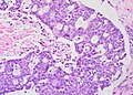
Histopathology [edit]
In case an ovarian cyst is surgically removed, a more than definite diagnosis tin be made by histopathology:
| Type | Subtype | Typical microscopy findings | Epitome |
|---|---|---|---|
| Functional cyst | Follicular cyst |
| 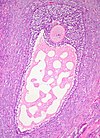 |
| Corpus luteum cyst |
| 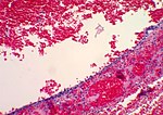 | |
| Cystadenoma | Serous cystadenoma | Cyst lining consisting of a simple epithelium, whose cells may be either:[21]
| 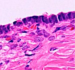 |
| Mucinous cystadenoma | Lined by a mucinous epithelium |  | |
| Dermoid cyst | Well-differentiated components from at least two germ layers (ectoderm, mesoderm and/or endoderm).[22] |  | |
| Endometriosis | At least two of the following iii criteria:[23]
|  | |
| Borderline tumor | Atypical epithelial proliferation without stromal invasion.[24] |  | |
| Ovarian cancer | Many different types, simply generally severe dysplasia/atypia and invasion. | Serous carcinoma. | |
| Simple squamous cyst | Simple squamous epithelium and non befitting to diagnoses above (a diagnosis of exclusion) |  | |
Treatment [edit]
Cysts associated with hypothyroidism or other endocrine bug are managed by treating the underlying status.
About 95% of ovarian cysts are beneficial, non cancerous.[25] Functional cysts and hemorrhagic ovarian cysts usually resolve spontaneously.[26] However, the bigger an ovarian cyst is, the less likely information technology is to disappear on its own.[27] Treatment may be required if cysts persist over several months, grow, or cause increasing pain.[28] Cysts that persist beyond ii or three menstrual cycles, or occur in post-menopausal women, may indicate more serious illness and should be investigated through ultrasonography and laparoscopy, particularly in cases where family members have had ovarian cancer. Such cysts may require surgical biopsy. Additionally, a blood test may exist taken before surgery to bank check for elevated CA-125, a tumour marking, which is often found in increased levels in ovarian cancer, although it can also exist elevated by other conditions resulting in a large number of fake positives.[29]
Hurting [edit]
Pain associated with ovarian cysts may be treated in several ways:
- Hurting relievers such equally acetaminophen, nonsteroidal anti-inflammatory drugs,[one] or opioids.
- While hormonal birth command prevents the development of new cysts in those who ofttimes get them,[1] it is non useful for the treatment of current cysts.[ii]
Surgery [edit]
Although most cases of ovarian cysts involve monitoring, some cases require surgery.[30] This may involve removing the cyst, or ane or both ovaries.[31] Technique is typically laparoscopic, unless the cyst is peculiarly large, or if pre-operative imaging suggests malignancy or complex anatomy.[32] In sure situations, the cyst is entirely removed, while with cysts with low recurrence adventure, younger patients, or which are in anatomically eloquent areas of the pelvis, they can be tuckered.[33] [34] Features that may indicate the demand for surgery include:[10]
- Persistent complex ovarian cysts
- Persistent cysts that are causing symptoms
- Complex ovarian cysts larger than 5 cm
- Simple ovarian cysts larger x cm or larger than 5 cm in postmenopausal patients
- Women who are menopausal or perimenopausal
Frequency [edit]
Most women of reproductive historic period develop small cysts each month, and large cysts that crusade bug occur in virtually 8% of women before menopause.[i] Ovarian cysts are nowadays in nearly 16% of women after menopause and if present are more likely to be cancer.[1] [3]
Benign ovarian cysts are common in asymptomatic premenarchal girls and found in approximately 68% of ovaries of girls 2–12 years sometime and in 84% of ovaries of girls 0–2 years old. Well-nigh of them are smaller than 9 mm while about 10–20% are larger macrocysts. While the smaller cysts mostly disappear within 6 months the larger ones appear to be more persistent.[35] [36]
References [edit]
- ^ a b c d eastward f m h i j k l m n o p q r s t u v w x y z aa ab ac ad ae "Ovarian cysts". Function on Women's Wellness. April 2019. Archived from the original on 12 August 2021.
- ^ a b Grimes, David A; Jones, LaShawn B.; Lopez, Laureen M; Schulz, Kenneth F (29 April 2014). "Oral contraceptives for functional ovarian cysts". Cochrane Database of Systematic Reviews (iv): CD006134. doi:x.1002/14651858.CD006134.pub5. PMID 24782304.
- ^ a b Mimoun, C.; Fritel, X.; Fauconnier, A.; Deffieux, X.; Dumont, A.; Huchon, C. (December 2013). "Épidémiologie des tumeurs ovariennes présumées bénignes" [Epidemiology of presumed beneficial ovarian tumors]. Journal de Gynécologie Obstétrique et Biologie de la Reproduction (in French). 42 (8): 722–729. doi:10.1016/j.jgyn.2013.09.027. PMID 24210235.
- ^ a b c d e f Ovarian Cysts at eMedicine
- ^ Legendre, Guillaume; Catala, Laurent; Morinière, Catherine; Lacoeuille, Céline; Boussion, Françoise; Sentilhes, Loïc; Descamps, Philippe (March 2014). "Relationship between ovarian cysts and infertility: what surgery and when?". Fertility and Sterility. 101 (3): 608–614. doi:ten.1016/j.fertnstert.2014.01.021. PMID 24559614.
- ^ Ovarian Cyst Rupture at eMedicine
- ^ "Ovarian cysts". womenshealth.gov. 2017-02-22. Retrieved 2020-10-29 .
- ^ "Ovarian Cysts Causes, Symptoms, Diagnosis, and Handling". eMedicineHealth.com. Archived from the original on 2007-03-07.
- ^ Abduljabbar, Hassan S.; Bukhari, Yasir A.; Hachim, Estabrq 1000. Al; Ashour, Ghazal Due south.; Amer, Afnan A.; Shaikhoon, Mohammed Chiliad.; Khojah, Mohammed I. (July 2015). "Review of 244 cases of ovarian cysts". Saudi Medical Journal. 36 (7): 834–838. doi:10.15537/smj.2015.7.11690. PMC4503903. PMID 26108588.
- ^ a b MedlinePlus Encyclopedia: Ovarian cysts
- ^ a b c Levine, Deborah; Brownish, Douglas Fifty.; Andreotti, Rochelle F.; Benacerraf, Beryl; Benson, Carol B.; Brewster, Wendy R; Coleman, Beverly; DePriest, Paul; Doubilet, Peter Thousand.; Goldstein, Steven R.; Hamper, Ulrike M.; Hecht, Jonathan 50.; Horrow, Mindy; Hur, Hye-Chun; Marnach, Mary; Patel, Maitray D.; Platt, Lawrence D.; Puscheck, Elizabeth; Smith-Bindman, Rebecca (September 2010). "Management of Asymptomatic Ovarian and Other Adnexal Cysts Imaged at U.s.: Society of Radiologists in Ultrasound Consensus Briefing Statement". Radiology. 256 (3): 943–954. doi:10.1148/radiol.10100213. PMC6939954. PMID 20505067. S2CID 10270209.
- ^ Kaijser, Jeroen; Sayasneh, Ahmad; Van Hoorde, Kirsten; Ghaem-Maghami, Sadaf; Bourne, Tom; Timmerman, Dirk; Van Calster, Ben (1 May 2014). "Presurgical diagnosis of adnexal tumours using mathematical models and scoring systems: a systematic review and meta-analysis". Homo Reproduction Update. xx (3): 449–462. doi:10.1093/humupd/dmt059. PMID 24327552.
- ^ Durbin, Kaci L.; Diaz-Montes, Teresa; Loveless, Meredith B. (August 2011). "Van Wyk and Grumbach Syndrome: An Unusual Case and Review of the Literature". Journal of Pediatric and Adolescent Gynecology. 24 (4): e93–e96. doi:10.1016/j.jpag.2010.08.003. PMID 21600802.
- ^ Dewailly, D.; Lujan, K. E.; Carmina, E.; Cedars, Thousand. I.; Laven, J.; Norman, R. J.; Escobar-Morreale, H. F. (1 May 2014). "Definition and significance of polycystic ovarian morphology: a task force report from the Androgen Excess and Polycystic Ovary Syndrome Order". Human Reproduction Update. 20 (iii): 334–352. doi:ten.1093/humupd/dmt061. PMID 24345633.
- ^ Altinkaya, Sunduz Ozlem; Talas, Betul Bayir; Gungor, Tayfun; Gulerman, Cavidan (October 2009). "Treatment of clomiphene citrate-related ovarian cysts in a prospective randomized study. A unmarried middle experience". Journal of Obstetrics and Gynaecology Enquiry. 35 (5): 940–945. doi:10.1111/j.1447-0756.2009.01041.x. PMID 20149045. S2CID 36836406.
- ^ Suzuki, Hirotada; Matsubara, Shigeki; Uchida, Shin-ichiro; Ohkuchi, Akihide (October 2014). "Ovary hyperstimulation syndrome accompanying molar pregnancy: case report and review of the literature". Athenaeum of Gynecology and Obstetrics. 290 (4): 803–806. doi:10.1007/s00404-014-3319-0. PMID 24966119. S2CID 27120087.
- ^ Nice clinical guidelines Issued: April 2011. Guideline CG122. Ovarian cancer: The recognition and initial direction of ovarian cancer Archived 2013-09-22 at the Wayback Car, Appendix D: Take a chance of malignancy alphabetize (RMI I).
- ^ a b c d east Network, Scottish Intercollegiate Guidelines (2003). "EPITHELIAL OVARIAN CANCER Section three: DIAGNOSIS". Epithelial ovarian cancer : a national clinical guideline. Edinburgh: SIGN. ISBN978-1899893935. Archived from the original on 2013-09-22.
- ^ a b Mohiedean Ghofrani. "Ovary - nontumor - Nonneoplastic cysts / other - Follicular cysts". Pathology Outlines. Topic Completed: 1 August 2011. Revised: 5 March 2020
- ^ a b c Aurelia Busca, Carlos Parra-Herran. "Ovary - nontumor - Nonneoplastic cysts / other - Corpus luteum cyst (CLC)". Pathology Outlines. Topic Completed: ane November 2016. Revised: 5 March 2020
- ^ Shahrzad Ehdaivand, M.D. "Ovary tumor - serous tumors - Serous cystadenoma / adenofibroma / surface papilloma". Pathology Outlines. Topic Completed: 1 June 2012. Revised: five March 2020
- ^ Sahin, Hilal; Abdullazade, Samir; Sanci, Muzaffer (Apr 2017). "Mature cystic teratoma of the ovary: a cutting edge overview on imaging features". Insights into Imaging. 8 (2): 227–241. doi:10.1007/s13244-016-0539-ix. PMC5359144. PMID 28105559.
- ^ Aurelia Busca, Carlos Parra-Herran. "Ovary - nontumor - Nonneoplastic cysts / other - Endometriosis". Pathology Outlines. Topic Completed: 1 August 2017. Revised: five March 2020
- ^ Lee-may Chen, MDJonathan South Berek, Doctor, MMS. "Deadline ovarian tumors". UpToDate.
{{cite web}}: CS1 maint: multiple names: authors listing (link) This topic terminal updated: Feb 08, 2019. - ^ "Symptoms of ovarian cysts". 2017-10-23. Archived from the original on 2009-05-12. Retrieved 2009-05-06 .
- ^ V.T. (14 May 2014). Understanding Ovarian Cyst. V.T. pp. 25–. GGKEY:JTX84XQARW9. Archived from the original on 16 March 2015.
- ^ Edward I. Bluth (2000). Ultrasound: A Practical Approach to Clinical Problems. Thieme. p. 190. ISBN978-0-86577-861-0. Archived from the original on 2017-03-12.
- ^ Susan A. Orshan (2008). Maternity, Newborn, and Women's Wellness Nursing: Comprehensive Care Across the Lifespan . Lippincott Williams & Wilkins. pp. 161. ISBN978-0-7817-4254-ii.
- ^ MedlinePlus Encyclopedia: CA-125
- ^ Tamparo, Carol; Lewis, Marcia (2011). Diseases of the Human Trunk. Philadelphia, PA: Library of Congress. p. 475. ISBN978-0-8036-2505-1.
- ^ "HealthHints: Gynecologic Health (Jan/February, 2003)". Texas AgriLife Extension Service: HealthHints. Archived from the original on 2007-02-17.
- ^ Surgit, O.; Gumus, I. Inegol (January 2014). "Unmarried-port Laparoscopic Total Hysterectomy and Bilateral Salpingooopherectomy Combined with Burch Colposuspension". Acta Chirurgica Belgica. 114 (four): 271–275. doi:10.1080/00015458.2014.11681025. PMID 26021429. S2CID 29262228.
- ^ Cho, Min Jeng; Kim, Dae Yeon; Kim, Seong Chul (October 2015). "Ovarian Cyst Aspiration in the Neonate: Minimally Invasive Surgery". Periodical of Pediatric and Adolescent Gynecology. 28 (five): 348–353. doi:10.1016/j.jpag.2014.10.003. PMID 26148782.
- ^ Nohuz, Due east. (January 2016). "Annotate je fais…fifty'évacuation d'un kyste annexiel sans ponctions itératives ni système d'aspiration-lavage lors d'une cœlioscopie" [How I exercise... the aspiration of an adnexal cyst without iterative needle punctures neither irrigation-aspiration device during a laparoscopy]. Gynécologie Obstétrique & Fertilité (in French). 44 (1): 63–66. doi:ten.1016/j.gyobfe.2015.11.001. PMID 26701109.
- ^ Cohen, H L; Eisenberg, P; Mandel, F; Haller, J O (July 1992). "Ovarian cysts are common in premenarchal girls: a sonographic study of 101 children two-12 years old". American Journal of Roentgenology. 159 (i): 89–91. doi:x.2214/ajr.159.1.1609728. PMID 1609728.
- ^ Qublan HS, Abdel-hadi J (2000). "Elementary ovarian cysts: Frequency and event in girls aged 2-9 years". Clinical and Experimental Obstetrics & Gynecology. 27 (i): 51–3. PMID 10758801.
Further reading [edit]
- McBee, W. C; Escobar, P. F; Falcone, T. (i February 2007). "Which ovarian masses need intervention?". Cleveland Clinic Journal of Medicine. 74 (ii): 149–157. doi:10.3949/ccjm.74.2.149. PMID 17333642.
- Simcock, B; Anderson, Due north (Feb 2005). "Diagnosis and management of simple ovarian cysts: An audit". Australasian Radiology. 49 (ane): 27–31. doi:10.1111/j.1440-1673.2005.01389.x. PMID 15727606.
- Ross, Elisa One thousand.; Kebria, Medhi (August 2013). "Incidental ovarian cysts: When to reassure, when to reassess, when to refer". Cleveland Dispensary Periodical of Medicine. lxxx (eight): 503–514. doi:10.3949/ccjm.80a.12155. PMID 23908107. S2CID 28081941.
- "Ovarian cyst - Treatment". National Health Service. 3 Oct 2018.
- Gerber, B.; Müller, H.; Külz, T.; Krause, A.; Reimer, T. (one Apr 1997). "Elementary ovarian cysts in premenopausal patients". International Journal of Gynecology & Obstetrics. 57 (1): 49–55. doi:x.1016/S0020-7292(97)02832-4. PMID 9175670. S2CID 34289061.
- Potter, Andrew Westward.; Chandrasekhar, Chitra A. (October 2008). "Usa and CT Evaluation of Acute Pelvic Pain of Gynecologic Origin in Nonpregnant Premenopausal Patients". RadioGraphics. 28 (6): 1645–1659. doi:x.1148/rg.286085504. PMID 18936027.
- Crespigny, Lachlan Ch.; Robinson, Hugh P.; Davoren, Ruth A. Yard.; Fortune, Denys (September 1989). "The 'simple' ovarian cyst: aspirate or operate?". BJOG. 96 (9): 1035–1039. doi:10.1111/j.1471-0528.1989.tb03377.ten. PMID 2679871. S2CID 22501317.
Source: https://en.wikipedia.org/wiki/Ovarian_cyst
0 Response to "If You Have Already Have Had a Ovarian Cyst Can You Get One Again"
Postar um comentário